
Brain Tumors Explained: Types, WHO Grades, and Multimodal Treatment Options
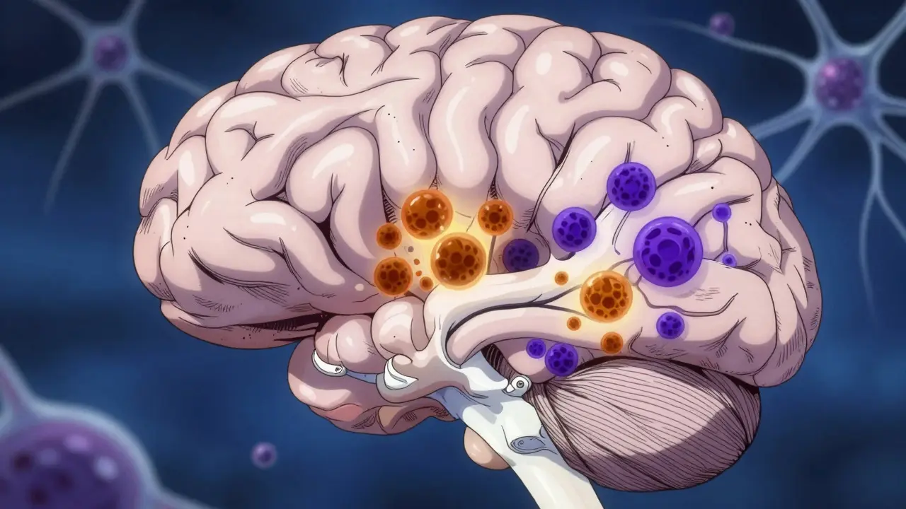
Receiving a brain tumor diagnosis can feel like the ground shifting under your feet. You’re suddenly confronted with medical jargon, urgent decisions, and a flood of information that often feels contradictory. Understanding the basics-what type of tumor you have, how aggressive it is, and what treatments are on the table-is the first step toward regaining some control.
Brain tumors are abnormal growths of cells in the central nervous system. They aren’t all the same. Some grow slowly and stay put; others race through tissue and spread. The difference matters because it dictates everything from surgery options to survival rates. In this guide, we’ll break down the types, the grading system used by doctors worldwide, and the modern multimodal approaches that combine surgery, radiation, and targeted drugs.
Key Takeaways
- Brain tumors are classified by both their cell type (histology) and molecular markers, not just how they look under a microscope.
- The World Health Organization (WHO) uses a 1-4 grading scale for most glial tumors, where Grade 1 is slow-growing and benign, while Grade 4 is highly aggressive and malignant.
- Molecular markers like IDH mutation status now determine treatment eligibility and prognosis more accurately than histology alone.
- Treatment is rarely one-size-fits-all; it typically involves a combination of neurosurgery, radiation therapy, and chemotherapy or targeted agents.
- Newer therapies, such as vorasidenib for specific low-grade gliomas, offer improved progression-free survival compared to traditional methods.
Understanding Tumor Types and Grading
Not all brain tumors are created equal. Doctors categorize them based on which cells they originate from. The most common group in adults is gliomas, which arise from glial cells that support neurons. Other types include meningiomas (growing from the meninges, the protective membranes around the brain), pituitary adenomas, and metastatic tumors that have spread from other parts of the body.
The critical part of the diagnosis is the grade. The WHO Classification of Tumors of the Central Nervous System, updated in its fifth edition (CNS5) in 2021, provides the global standard for this. For gliomas, grades range from 1 to 4:
- Grade 1: Slow-growing, well-defined edges, and rarely recur after removal. Pilocytic astrocytoma is a classic example.
- Grade 2: Low-grade but infiltrative. These tumors invade surrounding normal brain tissue and can eventually progress to higher grades. Diffuse astrocytoma falls here.
- Grade 3: Anaplastic tumors. These show rapid cell division and invasion. They are considered malignant and likely to recur.
- Grade 4: Highly aggressive. Glioblastoma is the prime example. It features necrotic (dead) centers, new blood vessel formation, and rapid growth.
A major shift in recent years is the integration of molecular data into this grading. For instance, an astrocytoma with an IDH mutation is treated differently than one without. The 2021 classification specifies that Oligodendroglioma must have both an IDH mutation and a 1p/19q codeletion to be diagnosed correctly. This precision helps predict how the tumor will behave and respond to treatment.
Diagnosis: Beyond the MRI Scan
An MRI scan gives a visual map of the tumor, showing its size, location, and whether it’s causing pressure on other areas. But imaging alone doesn’t tell you the full story. A biopsy is usually required to confirm the type and grade. During this procedure, a neurosurgeon removes a small sample of tissue for analysis.
Modern pathology labs don’t just look at cell shapes. They run a battery of tests to check for specific genetic changes. Key markers include:
- IDH1 and IDH2 mutations: Found in many lower-grade gliomas and associated with better outcomes.
- MGMT promoter methylation: Indicates how well a tumor might respond to temozolomide, a common chemotherapy drug.
- 1p/19q codeletion: Specific to oligodendrogliomas and linked to sensitivity to chemotherapy.
This comprehensive testing takes time. From biopsy to final diagnosis, patients should expect a wait of 7 to 10 business days. While waiting can be anxiety-inducing, these results are crucial. They transform a generic "brain tumor" label into a specific biological profile, allowing doctors to tailor the treatment plan precisely.
Standard Treatment Protocols
Treating brain tumors is almost always a team effort involving neurosurgeons, radiation oncologists, and neuro-oncologists. The goal is to remove as much tumor as safely possible, then use radiation and drugs to kill remaining cells.
Surgery
For accessible tumors, surgical resection is the first line of defense. The objective isn't always total removal, especially for infiltrative tumors that blend into healthy tissue. Instead, the aim is maximal safe resection-removing the visible tumor mass while preserving function. Advanced techniques like awake craniotomy (where the patient is awake during surgery to monitor speech and movement) and intraoperative MRI help surgeons navigate delicate areas.
Radiation Therapy
After surgery, radiation is often used to target microscopic disease left behind. Standard external beam radiation delivers high-energy beams over several weeks. Newer technologies like stereotactic radiosurgery (SRS) deliver a single, high dose of radiation to a precise spot, useful for smaller tumors or recurrent cases. For Grade 4 glioblastomas, radiation is combined with chemotherapy to maximize effectiveness.
Chemotherapy and Targeted Therapy
Drugs play a vital role, especially for tumors that cannot be fully removed. Temozolomide is the most common chemotherapy agent for gliomas. It works by damaging DNA in rapidly dividing cells. Its effectiveness depends heavily on MGMT methylation status; patients with methylated MGMT promoters tend to benefit significantly more.
Targeted therapies are also emerging. Vorasidenib, approved by the FDA in 2023, is a dual inhibitor of mutant IDH enzymes. It was specifically designed for Grade 2 gliomas with IDH mutations. Clinical trials showed it could double the time before the tumor progresses compared to placebo, offering a less toxic alternative to immediate chemotherapy for some patients.
Comparing Treatment Approaches by Grade
| Grade | Primary Goal | Common Interventions | Prognosis Notes |
|---|---|---|---|
| Grade 1 | Complete Removal | Surgery only; observation if residual remains | Cure is possible; recurrence is rare |
| Grade 2 | Maximal Safe Resection + Adjuvant Therapy | Surgery, Radiation, Chemotherapy (or Vorasidenib if IDH-mutant) | Long-term survival possible, but risk of progression exists |
| Grade 3 | Aggressive Control | Surgery, Radiation, Chemotherapy (Temozolomide) | Malignant; high risk of recurrence and transformation to Grade 4 |
| Grade 4 | Life Extension & Quality of Life | Surgery, Radiation + Concurrent Chemotherapy (Stupp Protocol) | Aggressive; median survival ~14-15 months with standard care |
Note that individual factors like age, overall health, and specific molecular markers can alter these general guidelines. A young patient with a Grade 2 oligodendroglioma may have a very different trajectory than an older adult with a Grade 3 astrocytoma.

Living with a Diagnosis: Practical Considerations
Beyond the medical procedures, managing a brain tumor diagnosis involves navigating emotional and logistical challenges. Many patients report feeling overwhelmed by the speed of decision-making. For example, fertility preservation needs to be addressed quickly before starting radiation or certain chemotherapies, which can impact reproductive health.
Support systems are crucial. Organizations like the National Brain Tumor Society provide resources, peer support groups, and updates on clinical trials. Joining a community can help you ask questions you might forget in the clinic and hear from others who have walked a similar path.
Keep a record of your symptoms, medications, and side effects. This log helps your medical team adjust treatments promptly. Also, don’t hesitate to seek a second opinion, especially for complex cases. Different institutions may have varying expertise in specific tumor subtypes or access to cutting-edge clinical trials.
Frequently Asked Questions
What is the difference between a benign and malignant brain tumor?
Benign tumors (usually Grade 1) grow slowly and have clear boundaries, making them easier to remove completely. Malignant tumors (Grades 3 and 4) grow rapidly, invade surrounding tissue, and are prone to recurring. However, even benign tumors can be dangerous if located in critical areas of the brain due to pressure effects.
How does the IDH mutation affect treatment?
An IDH mutation generally indicates a better prognosis and slower growth. It makes the tumor more sensitive to chemotherapy and radiation. Furthermore, it opens the door to targeted therapies like vorasidenib, which specifically inhibit the mutated enzyme. Without the mutation, treatment often relies more heavily on standard chemo-radiation protocols.
Is surgery always necessary for brain tumors?
Not always. If a tumor is deep within the brain or in a difficult-to-reach area, doctors might opt for radiation or chemotherapy first, or rely on active surveillance for very slow-growing lesions. The decision balances the benefit of removing tissue against the risk of neurological damage.
What is the Stupp Protocol?
The Stupp Protocol is the standard of care for Glioblastoma (Grade 4). It involves maximum safe surgical resection followed by concurrent radiation therapy and daily temozolomide chemotherapy, then continued maintenance temozolomide. This combination was established in 2005 and remains the baseline for treating this aggressive tumor.
How long does it take to get a final diagnosis after a biopsy?
Typically, it takes 7 to 10 business days. This period allows for detailed histological examination and molecular testing, including checks for IDH mutations and MGMT methylation. Rushing this process can lead to incomplete data, so patience is key for accurate treatment planning.